The report is the product
A label is not an answer. The system builds its case the way a careful morphologist would — count, measure, reason, show — and then writes it down, with its limits stated on the page. The full case is laid out below, section by section, with a note on why each step matters.
Peripheral blood — morphologic analysis
Specimen & method
- Input
- Digital cell gallery and whole-field image from a digital morphology analyser
- Scale calibration
- Self-derived from the erythrocyte population (median diameter ≡ 7.5 µm)
- Analysis
- Cell-type classification with conformal certification + morphometric census
- Clinical data
- None supplied — morphology only
Quantitative census
| Measure | Result | Basis |
|---|---|---|
| Nucleated cells | Direct enumeration | Segmented and counted, smudge cells excluded from the differential |
| Leucocyte : erythrocyte ratio | Markedly increased | vs ~1:700 in a normal film — a scale-free indicator |
| Dominant population | Small lymphoid cells | Single population, >85% of nucleated cells |
Morphometric findings
| Feature | Measured | Interpretation |
|---|---|---|
| Nuclear diameter | Small, low dispersion | Monomorphous — one dominant population rather than a reactive spectrum |
| Chromatin clumps / nucleus | 12 (median) | Coarsely clumped, condensed — a mature chromatin pattern |
| Internal chromatin SD | 20.9 | High contrast; blasts characteristically fall below 10 |
| Nuclear-to-cytoplasmic ratio | High | Scant cytoplasm |
| Smudge (Gumprecht) cells | Present | Detected by shape (circularity), not by paleness |
Morphologic impression
The appearances are those described in chronic lymphocytic leukaemia / small lymphocytic lymphoma (CLL/SLL).
Differential — and what the measurements exclude
| Considered | Status | On what basis |
|---|---|---|
| Reactive lymphocytosis | Against | Population is monomorphous; reactive expansions are pleomorphic with abundant cytoplasm |
| Acute lymphoblastic leukaemia | Excluded on chromatin | Blasts show finely dispersed chromatin with low internal SD; measured pattern is coarse and condensed |
| Mantle cell lymphoma | Not excluded | Cannot be separated on morphology alone — stated explicitly rather than omitted |
Recommended next steps are listed in the full report — for this pattern, flow-cytometric immunophenotyping is the discriminating test.
Cells of concern — annotated for review
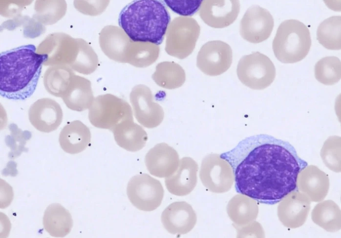
Representative field supplied by an expert hematopathology collaborator to illustrate the features named above; it is not an image of the case specimen.
Limitations — stated in every report
No clinical data or CBC accompanied this specimen; counts are areal estimates from the imaged field and carry sampling uncertainty. Morphology cannot separate CLL/SLL from mantle cell lymphoma. Where an automated count could not be supported by the image, the report says so rather than quoting a number — on this case the platelet estimate was invalidated on audit and reported as a floor.